HomeAbout UsContact Us
Dr. Manish Aggarwal offers advanced Endobronchial Ultrasound (EBUS) in Pitampura, Delhi for accurate, minimally invasive diagnosis of lung cancer, tuberculosis, sarcoidosis, enlarged lymph nodes, and other chest diseases through real-time ultrasound-guided evaluation and sampling.
Dr. Manish Aggarwal, Senior Interventional Pulmonologist & Chest Specialist in Pitampura, Delhi, offers advanced Endobronchial Ultrasound (EBUS) procedures for accurate diagnosis and staging of lung diseases, mediastinal lymph node enlargement, tuberculosis, sarcoidosis, and lung cancer.
With extensive experience in performing EBUS-guided procedures, the clinic provides minimally invasive real-time ultrasound-guided sampling of lymph nodes and deep chest lesions that are difficult to access through routine bronchoscopy. EBUS has become an essential tool in modern respiratory medicine for improving diagnostic accuracy while reducing the need for invasive surgical procedures.
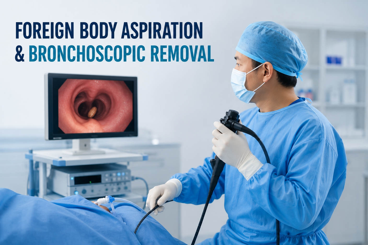
The procedure helps obtain larger and more accurate pleural tissue samples, significantly improving diagnostic yield in tuberculosis, cancer-related pleural disease, inflammatory disorders, and chronic pleural infections.
A complete range of advanced chest diagnostic services designed to evaluate lung nodules, enlarged lymph nodes, suspected cancer, sarcoidosis, tuberculosis, and other complex respiratory conditions.

Advanced endobronchial ultrasound-guided needle aspiration for accurate diagnosis and staging.

Comprehensive evaluation to determine the extent of lung cancer and guide treatment planning.

Minimally invasive lymph node sampling for diagnosis, staging, and treatment decisions.

Expert evaluation and tissue diagnosis for suspected sarcoidosis and related lung conditions.

Specialized assessment of tuberculosis-related lymph node enlargement using advanced diagnostics.

State-of-the-art bronchoscopic procedures for diagnosis and management of complex lung diseases.
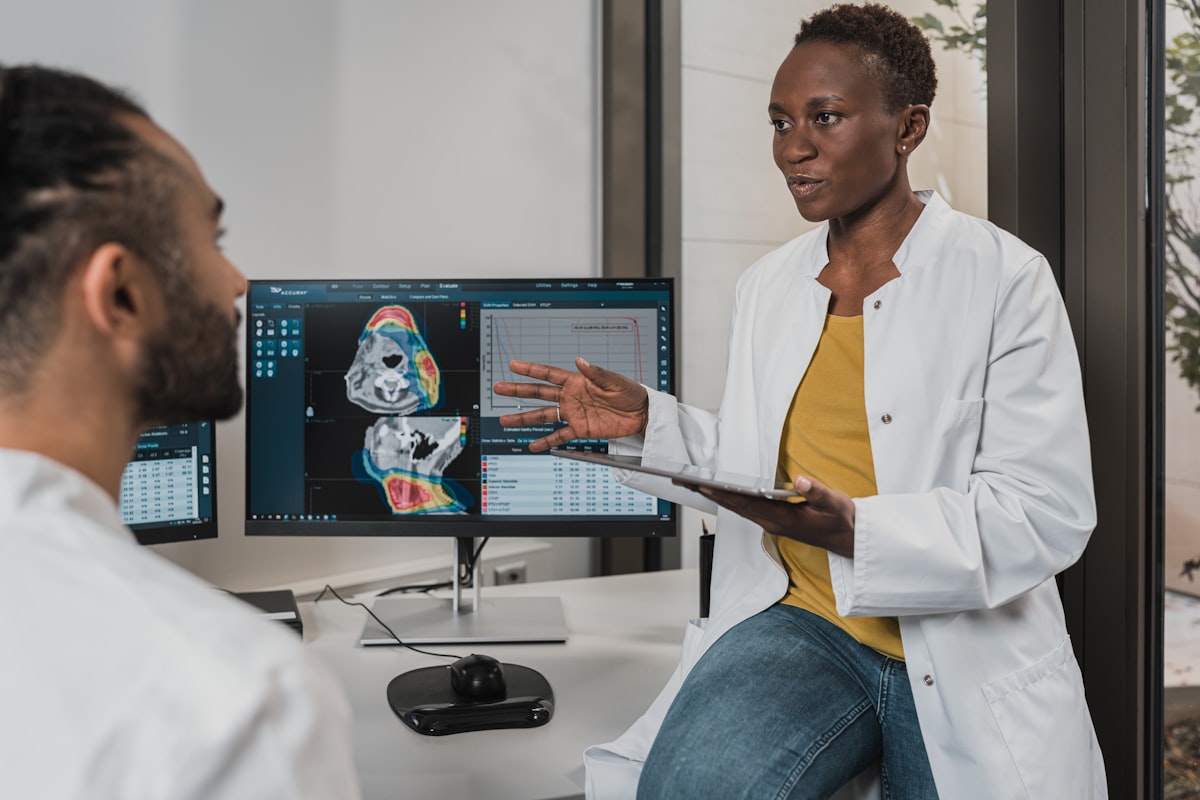
Precision diagnostic procedures designed to evaluate chest and lung conditions with minimal discomfort.

Comprehensive assessment of lung nodules and masses using advanced imaging and bronchoscopy.

Collaborative care involving pulmonology, oncology, radiology, and pathology specialists.
The clinic emphasizes patient safety, procedural precision, evidence-based diagnosis, and comprehensive respiratory care through advanced interventional pulmonology and minimally invasive bronchoscopic techniques.
Book consultation for expert EBUS evaluation and advanced chest disease diagnosis in Delhi.
Find answers to common questions about EBUS, EBUS-TBNA, lung cancer staging, mediastinal lymph node sampling, sarcoidosis diagnosis, tuberculosis evaluation, and advanced chest diagnostics at Delhi Lung & Bronchoscopy Center.
EBUS (Endobronchial Ultrasound) is an advanced bronchoscopic procedure that combines bronchoscopy with real-time ultrasound imaging. It allows pulmonologists to visualize and sample lymph nodes and structures located deep within the chest without the need for surgery.
EBUS-TBNA (Endobronchial Ultrasound-Guided Transbronchial Needle Aspiration) is a minimally invasive procedure that uses ultrasound guidance to obtain tissue samples from mediastinal lymph nodes and chest lesions for accurate diagnosis.
EBUS is commonly performed to evaluate enlarged chest lymph nodes, diagnose lung cancer, assess sarcoidosis, investigate tuberculosis, evaluate lung masses or nodules, and determine the cause of unexplained chest abnormalities.
EBUS allows precise sampling of lymph nodes and suspicious chest lesions, helping confirm the diagnosis of lung cancer and determine whether the disease has spread. This information is essential for accurate staging and treatment planning.
Yes. EBUS is frequently used to diagnose tuberculosis involving mediastinal lymph nodes, particularly when routine sputum tests are negative or when enlarged chest lymph nodes are present on imaging studies.
Yes. EBUS-TBNA is one of the most effective minimally invasive procedures for diagnosing sarcoidosis by obtaining tissue samples from enlarged mediastinal lymph nodes for pathological examination.
Mediastinal lymph nodes are lymph nodes located in the central part of the chest between the lungs. Enlargement of these lymph nodes can occur due to infections, tuberculosis, sarcoidosis, inflammation, or cancer.
EBUS may be recommended when CT scans or PET-CT scans reveal enlarged chest lymph nodes and a tissue diagnosis is required to determine the underlying cause.
In many situations, EBUS provides a safe and minimally invasive alternative to surgical procedures for obtaining tissue samples. It often reduces the need for more invasive diagnostic operations while maintaining high diagnostic accuracy.
Yes. EBUS can assist in evaluating selected lung nodules, masses, and lesions located near the airways, helping obtain tissue samples for diagnosis and treatment planning.
Most patients recover quickly after EBUS and can return home the same day, depending on the procedure performed and individual clinical circumstances. Recovery instructions are provided after the procedure.
Patients with enlarged chest lymph nodes, suspected lung cancer, sarcoidosis, tuberculosis, unexplained chest abnormalities, persistent lung lesions, or abnormal imaging findings may benefit from EBUS-guided evaluation.
We are available to answer queries, schedule urgent EBUS sessions or arrange Sleep diagnostic devices.