
HomeAbout UsContact Us
Explore documented diagnostic case files and clinical results under Dr. Manish Aggarwal. We combine advanced bronchoscopy, Level 1 sleep diagnostics, and NABL-compliant respiratory monitoring to treat common and complex lung diseases.

Years Experience
Bronchoscopies
EBUS Performed
Google Rating
Explore actual diagnostic pathways, procedural data, and outcome statistics managed by Dr. Manish Aggarwal at our East Delhi chest clinic.
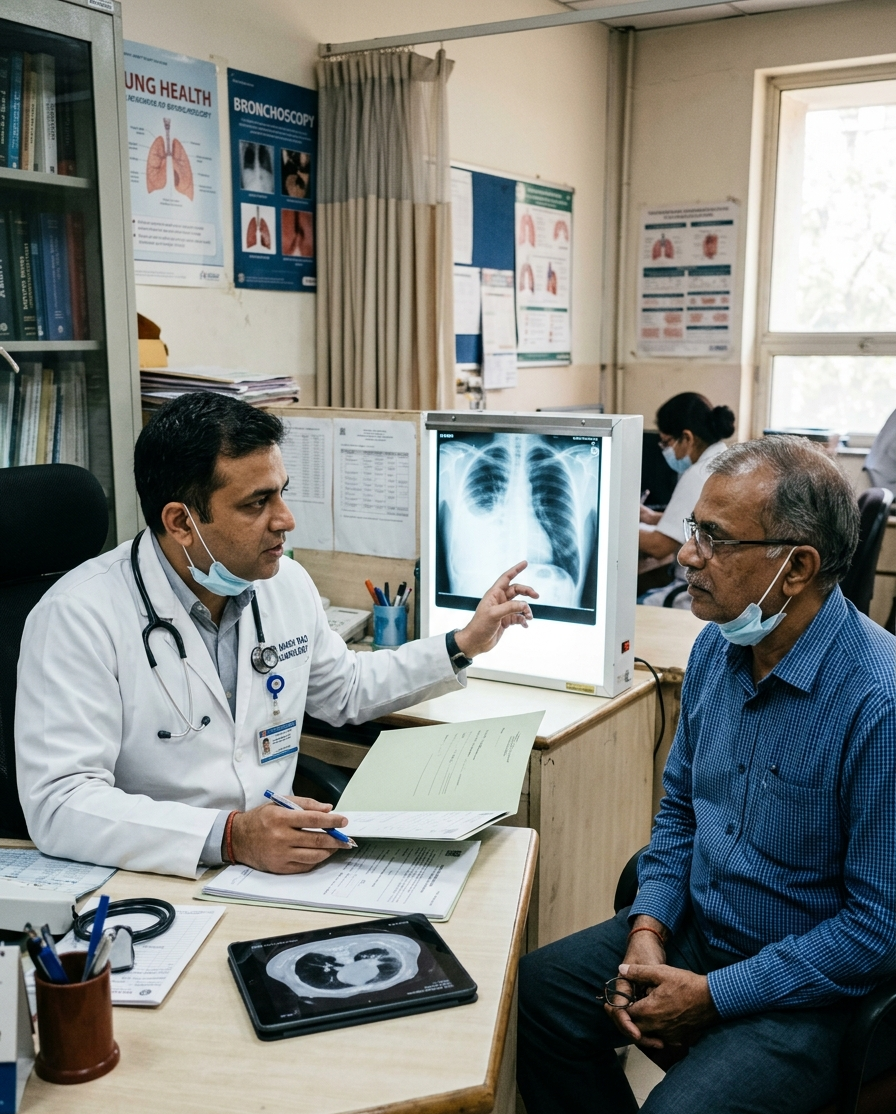 Full Case DetailInterventional Pulmonology
A 60-year-old gentleman came to our clinic with a persistent cough, increasing breathlessness, poor appetite, and a general sense of fatigue and ill health.
Malignant Pleural Effusion (Hiding within Pleural Tissue)
Sometimes, the diagnosis is not hidden because the tests were done incorrectly. Sometimes, it is hidden because the disease is waiting in a place that the tests never looked. And occasionally, finding the truth means looking beyond the fluid and directly at the tissue itself.
Quick Answer:
When fluid around the lungs keeps returning despite appropriate treatment for tuberculosis, it may be time to revisit the diagnosis. This real patient story highlights an important lesson in medicine: sometimes, the most valuable clue is not what tests show, but what happens when the patient fails to improve as expected.
Age
60 yrs
Gender
Male
Specialty
Interventional Pulmonology
A 60-year-old gentleman came to our clinic with a persistent cough, increasing breathlessness, poor appetite, and a general sense of fatigue and ill health.
A chest X-ray revealed a significant collection of fluid around his right lung, a condition known as pleural effusion. To understand this better, imagine the lung as a balloon sitting inside a thin protective pouch. When fluid accumulates within that pouch, it begins to compress the lung, making it difficult for it to expand fully during breathing.
A CT scan confirmed that the fluid had partially compressed the right lung. To investigate further, a bronchoscopy was performed. While there was no tumour visible within the airways, there appeared to be narrowing caused by pressure from outside the airway itself—an early clue that this case might not be straightforward.
In countries where tuberculosis (TB) is common, doctors frequently use a marker called ADA (Adenosine Deaminase) in pleural fluid to help identify patients likely to have TB-related pleural effusions. The pleural fluid was drained on three separate occasions, both to relieve his breathlessness and to establish the diagnosis. Each analysis appeared to support tuberculosis:
Based on initial supportive findings, initiating anti-tubercular treatment was a reasonable and evidence-based decision. At that stage, the diagnosis appeared settled. Unfortunately, the story was far from over.
Three weeks after starting appropriate anti-tubercular therapy, the patient returned with worsening breathlessness. The pleural fluid had accumulated again. And then again. The treatment response simply did not fit the expected pattern.
When the clinical picture and laboratory results fail to align, obtaining tissue directly from the pleura becomes essential. The patient underwent medical thoracoscopy, a minimally invasive procedure allowing direct visual examination of the lining surrounding the lungs.
Why Did the Fluid Tests Miss the Cancer?
Pleural fluid cytology detects cancer only when malignant cells are shed into the fluid in sufficient numbers. Some cancers remain confined predominantly to the pleural tissue and do not release many cells into the surrounding fluid.
Diagnostic Accuracy Performance Gap
Pleural fluid cytology may fail to detect cancer in approximately 40–50% of malignant pleural effusions. In such situations, a direct pleural biopsy obtained during thoracoscopy offers substantially higher accuracy.
The fluid was real. The elevated ADA was real. The inflammation was genuine. But the cancer itself was hiding within the pleural tissue rather than floating freely in the fluid. Multiple small abnormal nodules were identified over the pleural surface. Biopsies were obtained from these suspicious areas. The histopathology report provided the answer that three separate fluid analyses had failed to reveal. The underlying diagnosis was malignancy.
Although elevated ADA levels commonly occur in TB, they may occasionally be seen in certain cancers, lymphomas, and other inflammatory conditions.
Repeatedly negative fluid tests should not prevent further evaluation when clinical suspicion remains high.
When a patient does not respond as expected to an appropriate treatment, it is important to reassess the diagnosis rather than simply extending therapy indefinitely.
Direct visualisation of the pleura, combined with targeted biopsy, remains one of the most valuable diagnostic tools in unexplained recurrent pleural effusions.
Can pleural fluid have a high ADA level without tuberculosis?
Yes. Although elevated ADA levels strongly support the diagnosis of tuberculosis in the appropriate clinical setting, other conditions—including certain cancers, lymphoma, empyema, and autoimmune diseases—can occasionally produce similar results.
If pleural fluid cytology is negative multiple times, can cancer still be present?
Yes. Pleural fluid cytology may miss malignancy because some tumours do not shed cells readily into the fluid. In patients with persistent or recurrent pleural effusions, additional investigations such as thoracoscopy and pleural biopsy may still be required.
What should be done if pleural fluid keeps returning despite TB treatment?
Rapid re-accumulation of pleural fluid despite appropriate anti-tubercular therapy should prompt a re-evaluation of the diagnosis. In many cases, thoracoscopy with pleural biopsy may be recommended to obtain a definitive diagnosis.
Is thoracoscopy a major surgery?
Medical thoracoscopy is a minimally invasive procedure performed through a small incision in the chest wall. It allows the pulmonologist to inspect the pleura directly and obtain precise tissue samples. Recovery is generally quicker than with traditional surgical approaches.
Were the TB medicines a mistake?
Not necessarily. In regions where tuberculosis is common, starting anti-tubercular treatment based on strong clinical and laboratory evidence is often appropriate. However, when the expected improvement does not occur, clinicians must be prepared to reconsider the diagnosis and investigate further.
"When the patient's story and the test results do not match, it is the story that deserves another listen. Good medicine requires more than ordering investigations. It requires remaining curious, questioning assumptions, and being willing to revisit an earlier conclusion when the patient's progress tells us something different."
Sometimes, the diagnosis is not hidden because the tests were done incorrectly. Sometimes, it is hidden because the disease is waiting in a place that the tests never looked. And occasionally, finding the truth means looking beyond the fluid and directly at the tissue itself.
— Dr. Manish Aggarwal, Delhi Lung & Sleep Centre
This case has been shared for patient education purposes. Certain details have been modified to protect patient confidentiality.
Dr. Manish Aggarwal · Senior Chest Physician & Interventional Pulmonologist · Delhi Lung & Sleep Centre
 Full Case DetailPulmonology
A 45-year-old woman with asthma developed gradually worsening breathlessness over several weeks. Activities such as climbing stairs and performing household chores became increasingly difficult, leaving her breathless and exhausted.
Idiopathic Pulmonary Arterial Hypertension (IPAH)
Although pulmonary arterial hypertension remains a serious condition, early diagnosis and specialised treatment led to significant improvement in symptoms, activity levels, and overall quality of life.
Quick Answer:
A 45-year-old woman with asthma developed progressively worsening breathlessness and severe hypoxaemia with an oxygen saturation of 78%. Despite treatment for a presumed asthma exacerbation, her condition failed to improve. Further investigations ultimately revealed Idiopathic Pulmonary Arterial Hypertension (IPAH), a rare disorder affecting the blood vessels of the lungs.
Age
45 yrs
Gender
Female
Specialty
Pulmonology
A 45-year-old woman with asthma developed gradually worsening breathlessness over several weeks. Activities such as climbing stairs and performing household chores became increasingly difficult, leaving her breathless and exhausted.
The patient had a history of asthma and obesity (BMI 29) and had remained reasonably stable for years. As her breathlessness progressed, she initially assumed it was related to worsening asthma, weight gain, or stress. Concerned by her persistent symptoms, she checked her oxygen saturation at home and found it to be 78%.
She sought medical attention and was initially treated for a presumed asthma exacerbation with steroids, antibiotics, and supportive therapy. Despite treatment, her oxygen levels remained persistently low. The lack of improvement raised concerns that another diagnosis might be responsible for her symptoms.
When she presented to our clinic, the combination of progressive breathlessness and persistent hypoxaemia prompted a more detailed evaluation. An echocardiogram suggested markedly elevated pressures within the lung circulation. She was admitted for comprehensive assessment, and further investigations were performed to identify the underlying cause.
The patient was initially treated for a presumed asthma exacerbation using steroids, antibiotics, and supportive therapy. However, her oxygen levels remained low and her symptoms persisted.
Persistent hypoxaemia and progressive breathlessness prompted further evaluation. Echocardiography raised suspicion of elevated pulmonary artery pressures, leading to comprehensive assessment.
Following confirmation of Idiopathic Pulmonary Arterial Hypertension through right heart catheterisation, specialised medications were initiated to reduce pressure within the lung blood vessels and support heart function. Treatment was adjusted during regular follow-up based on clinical response.
Oxygen Saturation Clue
Home pulse oximetry revealed an oxygen saturation of 78%, an important warning sign that could not be fully explained by a routine asthma flare-up.
Diagnostic Confirmation
Right heart catheterisation remains the gold standard investigation for diagnosing pulmonary arterial hypertension and guiding treatment decisions.
Over the following months, her breathlessness improved, her activity levels increased, and she regained confidence in carrying out daily activities. Although pulmonary arterial hypertension remains a serious condition, timely diagnosis and appropriate treatment substantially improved her quality of life.
Patients with asthma may develop entirely separate lung or heart-related conditions that require different treatment.
Low oxygen levels that do not improve with standard treatment should prompt clinicians to reconsider the diagnosis.
When symptoms persist despite appropriate therapy, additional evaluation may reveal an alternative underlying condition.
It confirms pulmonary arterial hypertension and provides critical information for treatment planning.
Can asthma cause low oxygen levels?
Severe asthma can lower oxygen levels, but persistent hypoxaemia despite treatment requires further evaluation.
What is pulmonary arterial hypertension?
Pulmonary arterial hypertension is a condition in which the blood vessels of the lungs become abnormally narrowed, increasing the workload on the heart.
Why is right heart catheterisation important?
Right heart catheterisation confirms the diagnosis of pulmonary arterial hypertension and helps guide treatment decisions.
Is pulmonary arterial hypertension treatable?
While it may not always be curable, specialised therapies can improve symptoms and quality of life.
"Not every episode of breathlessness in an asthma patient is caused by asthma. Sometimes, symptoms are clues pointing toward a completely different diagnosis. Listening carefully to those clues and investigating further when expected improvement does not occur can make all the difference."
Although pulmonary arterial hypertension remains a serious condition, early diagnosis and specialised treatment led to significant improvement in symptoms, activity levels, and overall quality of life.
— Dr. Manish Aggarwal, Delhi Lung & Sleep Centre
This case has been shared for patient education purposes. Certain details have been modified to protect patient confidentiality.
Dr. Manish Aggarwal · Senior Chest Physician & Interventional Pulmonologist · Delhi Lung & Sleep Centre
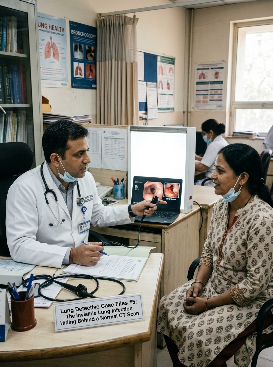 Full Case DetailInterventional Pulmonology
A 45-year-old woman presented with a persistent cough lasting nearly 120 days, accompanied by a low-grade intermittent fever. Despite multiple consultations and treatments, her symptoms continued to recur.
Endobronchial Tuberculosis (EBTB)
The patient responded well to Anti-Tubercular Therapy, with complete resolution of her chronic cough. Early diagnosis prevented irreversible airway scarring and bronchostenosis, which could have caused lifelong breathing difficulties.
Quick Answer:
A 45-year-old woman suffered from a persistent cough for nearly 120 days despite normal chest X-rays and CT scans. Further evaluation with flexible bronchoscopy revealed Endobronchial Tuberculosis (EBTB), a rare form of TB confined to the airway lining that can remain completely invisible on routine imaging.
Age
45 yrs
Gender
Female
Specialty
Interventional Pulmonology
A 45-year-old woman presented with a persistent cough lasting nearly 120 days, accompanied by a low-grade intermittent fever. Despite multiple consultations and treatments, her symptoms continued to recur.
The patient had undergone extensive evaluation including blood investigations, chest X-rays, and a Contrast-Enhanced Computed Tomography (CECT) scan of the chest and abdomen. All imaging studies appeared normal. Because no significant abnormality was identified, she was treated for chronic bronchitis and severe cough-variant asthma with multiple courses of antibiotics and oral steroids.
The oral steroids provided only temporary relief. Her cough would improve briefly before returning aggressively within days. This unusual response raised suspicion that the underlying cause was being masked rather than treated. The possibility of a hidden airway disease was considered despite repeatedly normal imaging.
Traditional radiological investigations failed to identify the source of her symptoms because the infection was confined entirely within the airway lining rather than the lung tissue itself. Recognising that the pathology might be hidden inside the bronchial tree, a flexible bronchoscopy was performed. Direct visualisation revealed a severely inflamed and ulcerated lesion obstructing the right main bronchus.
Based on normal imaging studies, the patient was treated as chronic bronchitis or cough-variant asthma using multiple courses of antibiotics and oral steroids. Symptoms improved only temporarily.
Because symptoms persisted despite treatment and radiological investigations remained normal, flexible bronchoscopy was performed to directly inspect the airway lining.
Following confirmation of Endobronchial Tuberculosis through biopsy and bronchial washings, the patient was started on standard weight-appropriate Anti-Tubercular Therapy (ATT).
Radiology Limitation
Standard chest X-rays and CT scans primarily evaluate lung tissue and may appear completely normal when Tuberculosis is confined exclusively to the airway lining.
Complication Prevention
Early diagnosis prevented progression to bronchostenosis, a permanent narrowing of the airway caused by chronic inflammation and scarring.
Within weeks of starting Anti-Tubercular Therapy, the patient's four-month-long cough resolved completely. The airway began healing, and the risk of permanent bronchial narrowing was avoided through timely intervention.
Endobronchial TB can remain entirely hidden when the infection is confined to the airway lining.
Recurrent symptoms after multiple courses of antibiotics and steroids should prompt reconsideration of the diagnosis.
Steroids may suppress inflammation and symptoms while allowing an underlying infection to continue progressing.
Direct visualisation of the airways allows identification of hidden infections, tumours, and structural abnormalities that imaging studies may miss.
Untreated Endobronchial Tuberculosis can lead to bronchostenosis, causing irreversible narrowing of the breathing tubes.
Can a chest CT scan miss Tuberculosis?
Yes. A chest CT scan can miss Tuberculosis if the infection is confined entirely to the airway lining, as occurs in Endobronchial Tuberculosis. Because CT scans primarily assess lung tissue, airway-limited disease may remain undetected.
What are the symptoms of Endobronchial Tuberculosis (EBTB)?
Common symptoms include a persistent cough lasting more than 3 to 4 weeks, low-grade intermittent fever, wheezing localized to one side of the chest, and shortness of breath that does not respond to standard asthma treatment or antibiotics.
Why does bronchoscopy detect hidden lung infections?
Flexible bronchoscopy allows direct visual inspection of the airway lining using a camera. It can identify hidden ulcers, infections, and growths while also allowing tissue biopsy for definitive diagnosis.
Why did the steroids help temporarily?
Steroids reduced airway inflammation caused by the infection, temporarily relieving symptoms. However, they did not treat the underlying Tuberculosis and may suppress the body's immune response against the infection.
What could have happened if the diagnosis was delayed?
Delayed diagnosis could have resulted in bronchostenosis, a permanent narrowing of the airway caused by chronic inflammation and scarring, leading to long-term breathing difficulties and possible lung collapse.
"Not every lung infection lives within the lung tissue itself. Sometimes the disease hides inside the airway lining where conventional imaging cannot see it. When symptoms persist despite normal scans, it is important to investigate beyond radiology and look directly at the airways."
The patient responded well to Anti-Tubercular Therapy, with complete resolution of her chronic cough. Early diagnosis prevented irreversible airway scarring and bronchostenosis, which could have caused lifelong breathing difficulties.
— Dr. Manish Aggarwal, Delhi Lung & Sleep Centre
This case has been shared for patient education purposes. Certain details have been modified to protect patient confidentiality.
Dr. Manish Aggarwal · Senior Chest Physician & Interventional Pulmonologist · Delhi Lung & Sleep Centre
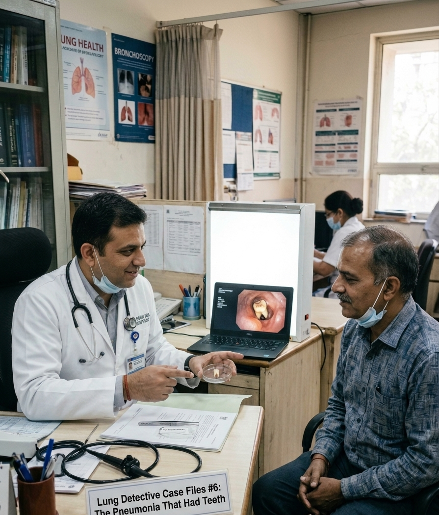 Full Case DetailInterventional Pulmonology
A 54-year-old man presented with a persistent productive cough, chest rattling, and a non-resolving pneumonia that had persisted for months despite multiple courses of antibiotics.
Post-Obstructive Pneumonia Secondary to Occult Foreign Body Aspiration (Human Tooth)
Removal of the impacted tooth restored normal airway function, cleared the chronic infection, resolved the patient's cough, and prevented serious complications such as lung abscess formation and permanent lung damage.
Quick Answer:
A 54-year-old man developed a non-resolving pneumonia that failed to improve despite multiple courses of antibiotics. Flexible bronchoscopy ultimately revealed a hidden tooth lodged within the right lower lobe bronchus. Removal of the foreign body cured the post-obstructive pneumonia and allowed the lung to heal.
Age
54 yrs
Gender
Male
Specialty
Interventional Pulmonology
A 54-year-old man presented with a persistent productive cough, chest rattling, and a non-resolving pneumonia that had persisted for months despite multiple courses of antibiotics.
The patient had undergone repeated treatment with broad-spectrum antibiotics for a localized pneumonia affecting the right lower lobe. Despite treatment, his symptoms continued and follow-up chest X-rays and CT scans showed a persistent area of inflammation that remained unchanged over time.
Multiple antibiotic regimens failed to provide meaningful improvement. The persistent lung shadow and ongoing symptoms suggested that a simple infection was unlikely to be the entire explanation. The possibility of an underlying mechanical airway obstruction was considered, as bacteria continued to thrive despite appropriate antimicrobial therapy.
A flexible bronchoscopy was performed to directly examine the airways. Deep within the right lower lobe bronchus, an unexpected foreign body was discovered. Hidden beneath inflamed tissue was a calcified object completely obstructing the airway. The object was identified as a human tooth, resulting in post-obstructive pneumonia caused by occult foreign body aspiration.
The patient received multiple courses of broad-spectrum antibiotics for presumed persistent bacterial pneumonia. Despite treatment, the infection failed to resolve.
When imaging findings remained unchanged and symptoms persisted, flexible bronchoscopy was performed to directly inspect the airway and identify a possible obstruction.
A specialized rat-tooth forceps was passed through the bronchoscope and used to securely grasp and remove the impacted tooth, eliminating the mechanical blockage and allowing the lung to recover.
Post-Obstructive Pneumonia Mechanism
When an airway becomes blocked, mucus clearance and normal airflow stop, creating an environment where bacteria can persist despite antibiotic therapy.
Lung Preservation Benefit
Early foreign body removal prevents complications such as lung abscess formation, tissue destruction, chronic infection, and permanent lung damage.
Following successful bronchoscopic extraction, the patient's airway reopened and normal drainage was restored. Within weeks, the chronic infection resolved completely, the cough disappeared, and follow-up imaging demonstrated significant clearing of the previously persistent lung shadow.
Mechanical airway obstruction can prevent a lung infection from resolving even when appropriate antibiotics are prescribed.
A localized pneumonia that repeatedly occurs in the same area should raise suspicion for an underlying blockage.
Teeth and other objects may enter the airway during sleep, dental procedures, trauma, or intoxication without the patient realizing it.
Direct airway visualization allows pulmonologists to identify hidden obstructions and remove them during the same procedure.
Delayed removal of airway foreign bodies can result in lung abscesses, chronic infection, tissue destruction, and permanent respiratory impairment.
Can you inhale a tooth into your lung without realizing it?
Yes. Occult foreign body aspiration is a recognized medical condition in adults and may occur during sleep, dental procedures, alcohol intoxication, sedation, or minor trauma without the patient being aware of the event.
What are the symptoms of a hidden foreign object in the lung?
Common symptoms include chronic cough, recurrent pneumonia affecting the same area of the lung, localized wheezing, chest rattling, and shortness of breath that does not improve with standard treatment.
Why didn't the antibiotics cure the pneumonia?
Although antibiotics can reduce bacterial growth, they cannot remove a physical obstruction. As long as the airway remains blocked, bacteria can continue to accumulate and cause persistent infection.
How do pulmonologists remove objects from the airway safely?
Pulmonologists use flexible or rigid bronchoscopy to visualize the airway and deploy specialized retrieval instruments such as baskets, graspers, or rat-tooth forceps to safely remove the foreign body.
What could happen if the foreign body remains untreated?
Untreated airway foreign bodies can lead to recurrent infections, lung abscesses, airway damage, tissue destruction, and permanent loss of lung function.
"When pneumonia remains fixed in the same location despite appropriate treatment, it is important to look beyond infection alone. Sometimes the problem is not the bacteria themselves but a hidden obstruction preventing the lung from healing. In these situations, bronchoscopy often provides the answer."
Removal of the impacted tooth restored normal airway function, cleared the chronic infection, resolved the patient's cough, and prevented serious complications such as lung abscess formation and permanent lung damage.
— Dr. Manish Aggarwal, Delhi Lung & Sleep Centre
This case has been shared for patient education purposes. Certain details have been modified to protect patient confidentiality.
Dr. Manish Aggarwal · Senior Chest Physician & Interventional Pulmonologist · Delhi Lung & Sleep Centre
Tap "Read Full Case" to explore detailed clinical files
Breathe better, sleep well. Dr. Manish Aggarwal, pulmonologist & sleep specialists deliver world-class treatment, right here in Pitampura (Currently in Shalimar Bagh), Delhi.
17+
Specialties
50k+
Patients Treated
24 yr+
Experience

Pulmonology
Advanced Care
Chronic Sleep
Advanced Treatment
Book An Appointment Today
Get instant answers to common queries regarding chest diagnostics, sleep assessments, and clinical parameters at Delhi Lung & Bronchoscopy Center.
This case study provides a detailed overview of the client's challenges, our strategic approach, the implementation process, and the measurable results achieved.
Performance was tracked using key metrics such as website traffic, lead generation, conversion rates, engagement, and overall business growth over the project duration.
Yes. All figures and performance indicators presented in this case study are based on actual project data and verified campaign analytics.
Every business has unique goals and market conditions. While strategies can be adapted, actual results may vary depending on competition, budget, and implementation.
The timeline varies by project, but most measurable improvements are observed within the first few months after implementing the recommended strategies.
Our case studies span healthcare, digital marketing, local businesses, eCommerce, technology, and professional service industries.
Absolutely. Our team can evaluate your current challenges and develop a customized strategy tailored to your industry and growth objectives.
You can contact our team through the inquiry form or schedule a consultation to discuss your project requirements and business goals.
We are available to answer queries, schedule urgent EBUS sessions or arrange Sleep diagnostic devices.